Quantitative Analysis of Oxytocin Receptor Densities in the Brain of the Coyote (Canis Latrans)
Research Introduction
- Oxytocin is a neuropeptide that has been shown to be a factor in species that display social monogamy1. The neural actions of oxytocin at the oxytocin receptor in the brain are necessary for social memory of familiar individuals of the same species.
- Much research has been done on oxytocin receptors (OXTR) in the brains of socially monogamous rodents and non-human primates, and these studies have demonstrated a critical role of oxytocin in the neurobiology of social attachment.
- Coyotes are a unique species in the context of social research because they are socially monogamous and have been shown to also exhibit sexual and genetic monogamy.
The goal of the current study is to quantify the distribution of OXTR throughout the coyote forebrain in order to compare their receptor map to other known monogamous species and to lay the neuroanatomical foundation for future studies of the oxytocin system of coyotes.
Methods
Animal Specimens
In order to map coyote OXTR, we utilized six brains that were opportunistically collected from captive-housed coyotes at the USDA Millville Predator Research Center. These animals were euthanized for other reasons unrelated to this study.
Specimen Preparation
The brain samples were fresh frozen on dry ice within hours of death and cut into blocks. The blocks were then sectioned at 20 microns using a cryostat, and the slices mounted on glass slides.
Receptor Autoradiography
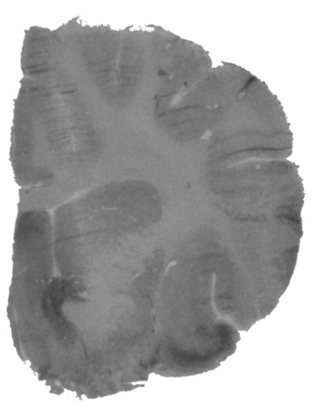
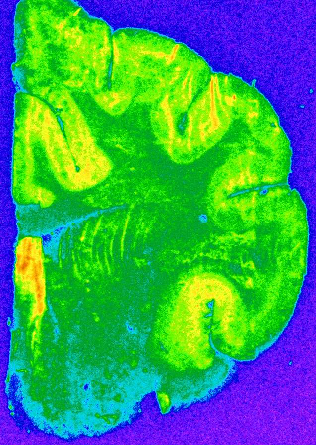
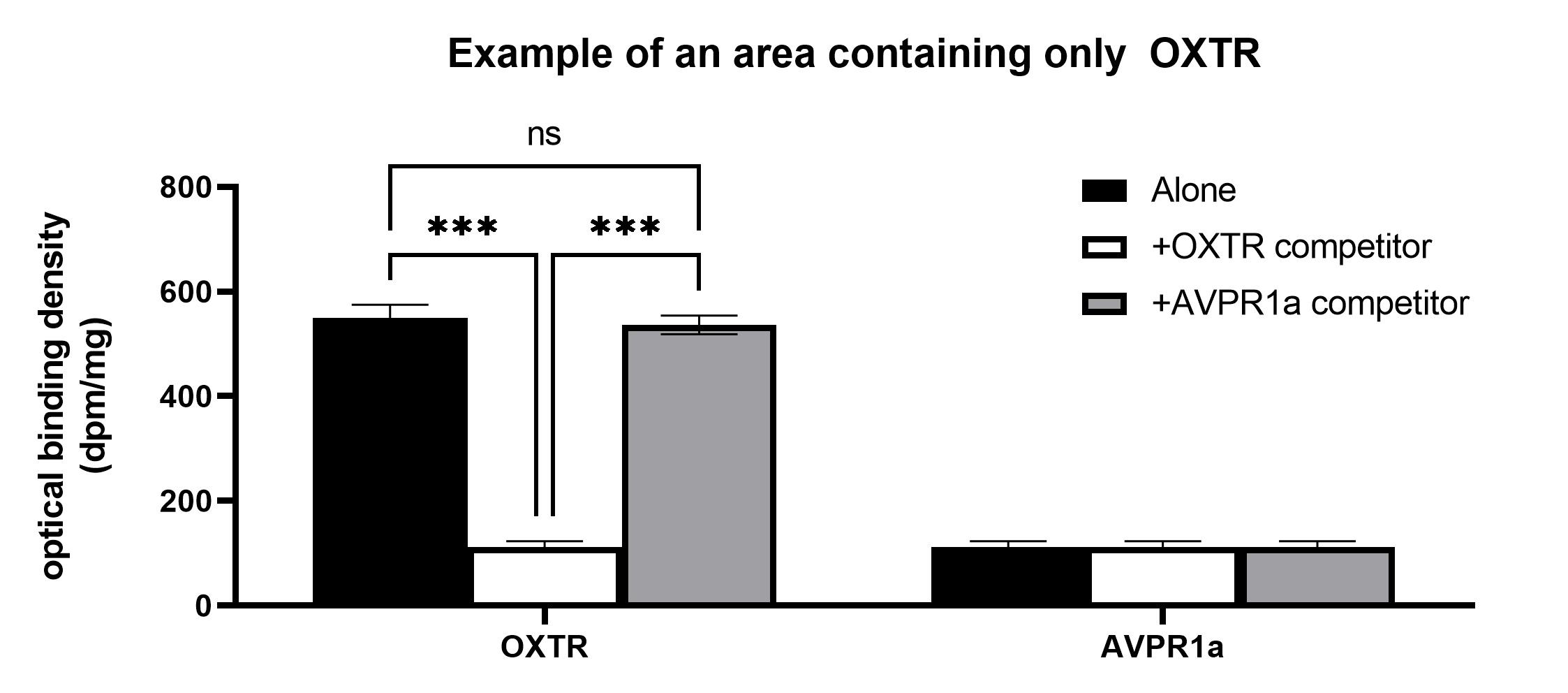
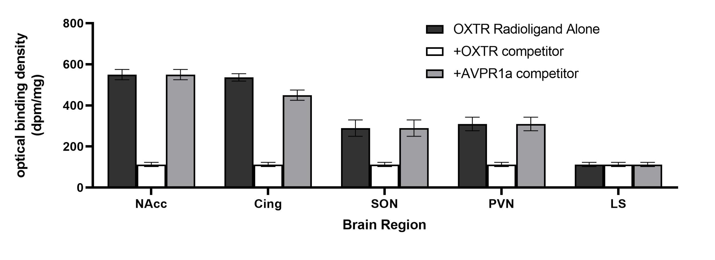
These slides were processed according to established methods to visualize OXTR using receptor autoradiography2. This process uses radioactively labeled ligands that bind to OXTR and emit radiation, which is detected by radiosensitive film (Figure 1 & 2). We will then take these films and quantify them by using digital densitometry (Figure 3). As part of the autoradiography procedures we will also use an OXTR and Vasopressin 1a Receptor inhibitor in order to insure that our ligand is binding to OXTR receptors (Figure 4 & 5).
Note: These results are hypothetical. We have not yet obtained data. Quantification is planned for Spring 2021.
Note: These results are hypothetical. We have not yet obtained data. Quantification is planned for Spring 2021.
Expected Results
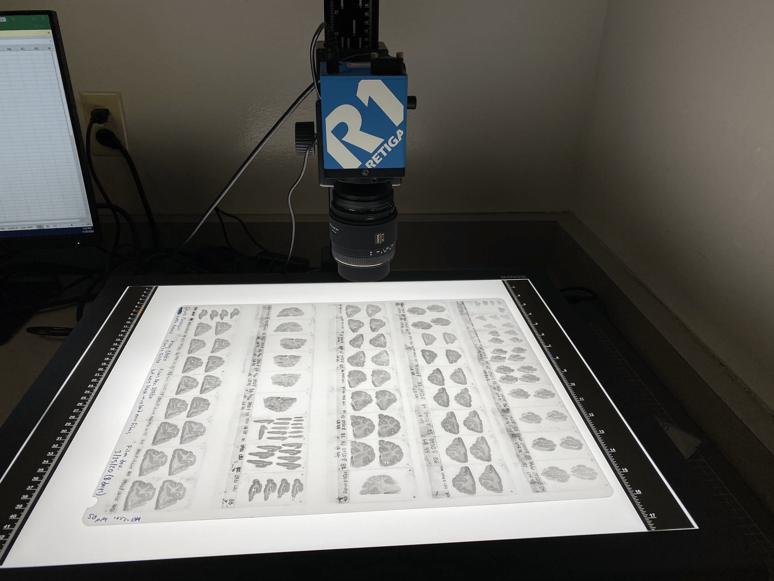
We will use a digital densitometry software package and a lightbox with a top mounted camera (Figure 1) to outline our regions of interest and measure the density of radioligand binding (i.e. receptor density).
Conclusions & Future Directions
- While previous studies were largely qualitative, these planned analyses will provide quantitative results, characterizing the oxytocin receptor distribution for the first time.
- If coyote oxytocin receptor distribution follows a pattern similar to those of other monogamous species, we will confirm our hypothesis and lend support to conserved oxytocin pathways convergently giving rise to monogamous behaviors in various mammalian species.
References
- Young, L. J. & Wang, Z. The neurobiology of pair bonding. Nature Neuroscience 7, 1048–1054 (2004).
- Freeman SM, Walum H, Inoue K, Smith AL, Goodman MM, Bales KL, Young LJ. Neuroanatomical distribution of oxytocin and vasopressin 1a receptors in the socially monogamous coppery titi monkey (Callicebus cupreus). Neuroscience 2014; 273: 12–23.
- Palazzi, Xavier; The Beagle Brain in Stereotaxic Coordinates; Springer Science & Business Media; 2011.