Distribution of Vasopressin 1a Receptor Binding in the Brain of Coyotes (Canis latrans)
Distribution of Vasopressin 1a Receptor Binding in the Brain of Coyotes (Canis latrans) PDF File
Introduction
Vasopressin
Known as Arginine Vasopressin (AVP), it is a neuropeptide hormone that plays a role in the biological basis of social attachment in monogamous species. AVP functions in the brain to modulate social memory, territoriality, and social attachment between pair-bonded adult mates of the same species.
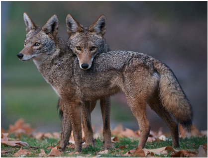
Coyotes
A unique species in the context of social research because they are socially monogamous and have been shown to also exhibit sexual and genetic monogamy, which is rare among mammals.
Our Goals
- Establish the distribution of vasopressin 1a receptors (AVPR1a) throughout the coyote forebrain in order to compare their receptor distribution map to other known monogamous species.
- Provide a neuroanatomical foundation for future studies of the AVP system of coyotes.
Predictions
Based off of published studies and our previous results from our last study, we predict that we will find receptors in the lateral septum, ventral pallidum, cingulate cortex, and hypothalamus.
Methods
Animals
Six brains (3 male, 3 female) were opportunistically collected from captive-housed coyotes at the USDA Millville Predator Research Center that were euthanized for reasons unrelated to the current study.
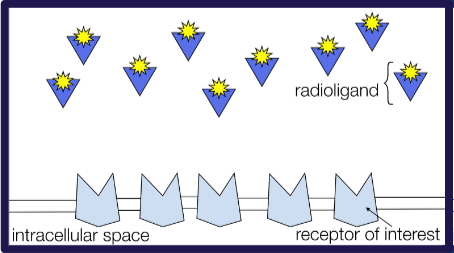
Specimen Preparation & Autoradiograpny
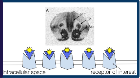
The samples were fresh frozen on dry ice within hours of death and sectioned at 20 μm using a cryostat. We processed them using AVPR1a autoradiography according to well established procedures1, which are shown in the schematic in Figure 1.
Analysis
We used a brain atlas from a beagle2, which as similar structure to the coyote, to identify the areas of brain that contain AVPR1a. Receptor binding was quantified using a digital densitometry system that was calibrated using ten radioactive microscales. Regional differences were determined using a one-way, repeated measures ANOVA with paired, post-hoc comparisons across all regions. Sex differences across regions were determined using a two-way, repeated measures ANOVA.
Results
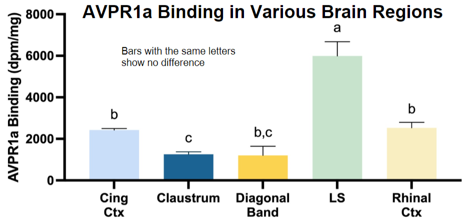
- We observed AVPR1a binding in two regions that have been previously shown to express these receptors in other social animals: the Diagonal Band (4A) and the Lateral Striatum (4C).
- We also found binding in the cerebral cortex, especially the Rhinal Cortex (4B) and the Cingulate Cortex (4D).
- We also detected binding in the Claustrum (4E).
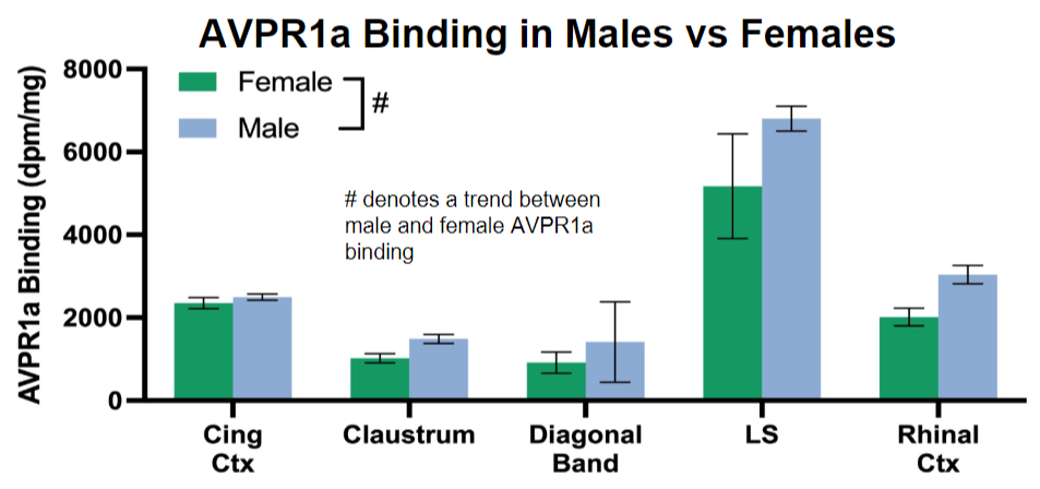
- The Lateral Septum had higher binding compared to all other brain regions (Fig 2).
- When comparing binding in males and females, no significant difference was found, but there was a trend (p=0.062) that males had higher AVPR1a binding than females across all regions (Fig 3).
Conclusions & Future Directions
- AVPR1a receptors are spread in various regions of the coyote brain, which is a similar patter to other monogamous species.
- AVPR1a is in areas involved in memory (Rhinal cortex).
- Our low sample size likely contributed to the trend of males having higher AVPR1a binding, which did not reach significance.
- Future directions include:
- counterstain sections to confirm anatomical boundaries.
- continue to section additional brains and deeper anatomical levels and compare to known receptor distributions in other monogamous mammals.
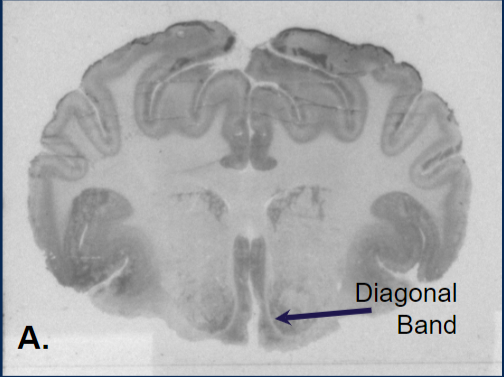
Image A
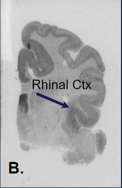
Image B
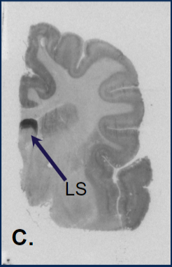
Image C
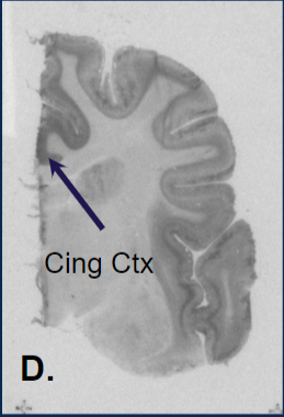
Image D
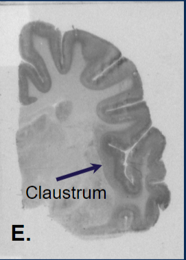
Image E
Images A-E are autoradiograms of the coyote brain tissue (Brain regions established using a beagle brain atlas2)
References
- Freeman SM, Walum H, Inoue K, et al. Neuroanatomical distribution of oxytocin and vasopressin 1a receptors in the socially monogamous coppery titi monkey (Callicebus cupreus). Neuroscience. 2014;273:12-23. doi:10.1016/j.neuroscience.2014.04.055
- Palazzi, Xavier. The Beagle Brain in Stereotaxic Coordinates. Springer, 2011