By Kayla Suisse, David Suisse, Dr. Amita Kaundal
Antimicrobial Activity of Artemisia tridentata
Introduction
- Plant secondary metabolite produced by plants may have the capability to inhibit the growth of microbes or to modify the plant's microbiome.
- These chemicals from various medicinal plants have been used for centuries to treat multiple medical conditions and diseases.
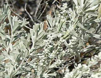
- Artemisia tridentata commonly referred to as "Big Sagebrush," is one such medicinal plant traditionally used by Native Americans to relieve stomach pain, colds and coughs, sore eyes, snake bites, and as an insect repellent.
- We are interested in studying the reported medicinal properties of A. tridentata, precisely its antimicrobial properties. This study tested crude extracts from the sagebrush's twigs, leaves, and flowers to identify their antimicrobial activities against various human and plant pathogens.
Figure 1: Artemisia tridentata
Methods
Preparation of Plant Crude Extracts
- Samples of A. tridentata leaves, twigs, and flowers were collected from various locations in Green Canyon.
- They were washed and dried for a month and ground to a fine powder. Methanol was used to create a concentrated solution. The solutions were then dried using a rotary evaporator.
Antimicrobial Assay of Plant Crude Extracts by Disk Diffusion Method
- The dried extracts were resuspended in Dimethyl Sulfoxide to 50mg/ml.
- Filter paper disks soaked in50μl (2.5mg) and 100μl (5mg) of leaf, twig and flower extract were tested against B. subtilis, E.coli Dh5α, Agrobacterium tumefaciens, Pseudomonas syringae pv DC3000, P.syringae pv tabaci, Proteus vulgaris, Staphylococcus aureus, S. epidimides, and Micrococcus luteus for growth inhibition on Mueller Hinton Agar.
- 10, 25, 30, and 50 μg ampicillin, gentamycin, streptomycin, vancomycin, and chloramphenicol disks used as positive control. Disk soaked in DMSO used as negative control.
- Plates were incubated for 12-16 hours in incubator and observed for zone of inhibition, which were then measured.
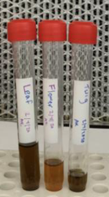
Figure 2: Crude Extract
Identification of Antimicrobial Compounds
- Samples of leaf and flower extract were separated into fractions through HPLC analysis and fractions were collected upon separation.
- Each fraction was evaporated onto filter paper disks and was tested against B. subtilis for antimicrobial activity.

(top) 100μL (5mg) flower disk (center) 50μL (2.5mg) flower well

(top) 100μL (5mg) leaf disk (center) 50μL (2.5mg) leaf well

(left) Fraction #4 disk
Flower
| 2.5mg well | 0.5mg disk | 2.5mg disk | 5.0mg disk | Ampicillin (10) | Ampicillin(50) | Gentamycin(50) | Streptomycin(50) | Kanamycin(10) | Kanamycin(50) | Chloramphenicol(30) | Vancomycin(30) | |
|---|---|---|---|---|---|---|---|---|---|---|---|---|
| Bacillus subtilis (mm) | 2.5 | ND | 1.5 | 5.0 | ND | 5.7 | 7.2 | 3.8 | ND | ND | ND | ND |
| Agrobacterium tumefaciens (mm) | 2.0 | ND | 1.0 | 3.0 | ND | 0 | 0 | 0 | 0 | ND | ND | ND |
| P. syringae pv tabaci (mm) | 2.7 | 0 | 0 | ND | 2.2 | ND | ND | ND | ND | ND | ND | ND |
| Escherichia coli Dh5a (mm) | 1.0 | 0 | 0 | ND | 0 | 1.1 | 3.3 | ND | 5.0 | 5.6 | ND | ND |
| Bacillus cereus (mm) | ND | ND | ND | 4.0 | ND | 1.0 | 9.2 | ND | ND | ND | ND | ND |
| Proteus vulgaris (mm) | ND | ND | ND | 0.5 | ND | 0.0 | ND | ND | ND | ND | ND | ND |
| Micrococcus luteus (mm) | ND | ND | ND | 7.0 | ND | ND | ND | ND | ND | ND | ND | 9.0 |
| Staphylococcus aureus (mm) | ND | ND | ND | 1.5 | ND | ND | ND | ND | ND | ND | 1.5 | ND |
| Staphylococcus epidimides (mm) | ND | ND | ND | 3.0 | ND | ND | ND | ND | ND | ND | 3.0 | ND |
Twig
| 2.5mg well | 0.5mg disk | 2.5mg disk | 5.0mg disk | Ampicillin (10) | Ampicillin(50) | Gentamycin(50) | Streptomycin(50) | Kanamycin(10) | Kanamycin(50) | Chloramphenicol(30) | Vancomycin(30) | |
|---|---|---|---|---|---|---|---|---|---|---|---|---|
| Bacillus subtilis (mm) | 4.0 | 0 | 1.1.0 | 5.0 | ND | 5.7 | 7.2 | 3.8 | ND | ND | ND | ND |
| Agrobacterium tumefaciens (mm) | 3.0 | ND | 2.0 | 1.0 | ND | 0 | 0 | 0 | 0 | 0 | ND | ND |
| P. syringae pv tabaci (mm) | 3.3 | ND | 0 | ND | 2.2 | ND | ND | ND | ND | ND | ND | ND |
Leaf
| 2.5mg well | 0.5mg disk | 2.5mg disk | 5.0mg disk | Ampicillin (10) | Ampicillin(50) | Gentamycin(50) | Streptomycin(50) | Kanamycin(10) | Kanamycin(50) | Chloramphenicol(30) | Vancomycin(30) | |
|---|---|---|---|---|---|---|---|---|---|---|---|---|
| Bacillus subtilis (mm) | 2.0 | 2.0 | 1.0 | 2.0 | ND | 5.7 | 7.2 | 3.8 | ND | ND | ND | ND |
| Agrobacterium tumefaciens (mm) | 2.0 | ND | 1.0 | 3.0 | ND | 0 | 0 | 0 | 0 | ND | ND | ND |
| P. syringae pv tabaci (mm) | 2.7 | 0 | 0 | ND | 2.2 | ND | ND | ND | ND | ND | ND | ND |
| Bacillus cereus (mm) | ND | ND | ND | 3.5 | ND | 1.0 | 9.2 | ND | ND | ND | ND | ND |
| Proteus vulgaris (mm) | ND | ND | ND | 0.5 | ND | 0.0 | ND | ND | ND | ND | ND | ND |
| Micrococcus luteus (mm) | ND | ND | ND | 5.0 | ND | ND | ND | ND | ND | ND | ND | 9.0 |
| Staphylococcus aureus (mm) | ND | ND | ND | 0.0 | ND | ND | ND | ND | ND | ND | 1.5 | ND |
| Staphylococcus epidimides (mm) | ND | ND | ND | 3.0 | ND | ND | ND | ND | ND | ND | 3.0 | ND |
- Preliminary results showed that the crude extract from all three parts of Artemisia tridentata showed antimicrobial activity.
- Flower extract showed the most potent antimicrobial activity.
- B. subtilis is more sensitive to the crude extract.
- Fraction 4, showed antimicrobial activity.
Future Plan
- Results of BSL 1 and 2 pathogen assays may be used to ascertain some aspects of the identity of antimicrobial components.
- We are working to separate Fraction 4 of the extract by GCMS to identify the compounds present in the fraction, as it was the only fraction to show antimicrobial activity in preliminary testing, per Figure 3c.