By Gentry Mower, Ariel Snowden, Dr. Sara Freeman
Oxytocin Receptors in Substantia Nigra of the Brain in Individuals with Schizophrenia
Introduction
- Schizophrenia (SZ) is a severe, chronic disorder, which affects about 2 million individuals in the United States alone1. SZ is characterized by both positive symptoms (hallucinations, delusions) and negative symptoms (cognitive deficits, impaired social cognition). Its health, social, and economic burden is tremendous2.
- Most antipsychotic drugs, targeting the positive symptoms of SZ, leave the social cognitive aspect the same or worsened3. Because the level of social cognition is the largest predictor of an individual’s functional independence, there is a huge need for better treatment options for individuals with SZ.
- Oxytocin is a hormone that mediates many complex social behaviors in both humans and animals. It is involved in maternal bonding, pair mate selection, and social cognition4. Studies identify intranasal oxytocin as a potential way to alleviate the social impairments observed in individuals with SZ5.
- Our aim is to localize the oxytocin receptors (OXTR) in the brains of individuals who had SZ—specifically in the substantial nigra (SN; Figure 1). Without a clear understanding of where the OXTR are located in the human brain, it is impossible to understand the mechanism of OXT on the brain in a neurotypical or clinical population. Preliminary work from the Freeman lab has identified SN as a region of interest. The SN is involved in the dopaminergic pathway, which is typically heightened in individuals with SZ. A statistically significant difference would imply that the SN is a target for using OXT as a treatment to alleviate the negative symptoms of SZ.
Methods
- Human tissue was obtained from the NIH NeuroBioBank. We received 16 control (7 F, 9 M) and 16 SZ (10 F, 6 M) specimens (Table 1). Each specimen was frozen at -80°C until sectioned at 20 microns using a cryostat. Slices were mounted on glass slides.
- These slides were processed according to previously established methods to visualize OXTR using receptor autoradiography 6. These methods employ competitive binding techniques, ultimately differentiating between regions of high OXT binding and vasopressin (AVP) in the brain, as these neuropeptide hormones are structurally similar. Radioactively labeled ligands bind to OXTR, and emitted radiation is detected by radiosensitive film. We then quantified the density of each region digitally.
- We employed two statistical tests: one-way repeated measure ANOVA between the three radioactive conditional groups to ensure that our results are not confounded by AVP binding. We also performed a post hoc paired samples t-test to reveal any significant differences in OBD between SZ and control groups.
Table 1: Tissue Received
| Schizophrenia | Unaffected Matched Controls | |
|---|---|---|
| Requested | N=22 | N=22 |
| Recieved | N=16 | N=16 |
| Females | 10 | 7 |
| Males | 6 | 9 |
| Age Range | 54-83 | 47-83 |
Figure 1
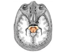
Location of the human substantia nigra.
Figure 2
Control
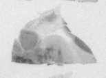
Control
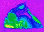
Schizophrenia
Schizophrenia
Slices of human brain after treatment with radioactive OXTR ligand alone. “Warmer” regions indicative of higher Optical Binding Density (OBD), indicating a larger density of OXTR in that region of brain.
Results
- We successfully visualized OXTR in the substantia nigra of our specimens (Figure 2, right).
- One way repeated measure ANOVA showed significant effect of condition on OXTR binding between OXTR alone relative to +1nM AVPR1a-A and +100nm OXTR-A (Figure 3).
- A post hoc unpaired t-test revealed no significant difference in OXTR OBD between SZ (5824 dpm/mg) and Control (5485 dpm/mg), p=0.6796 (Figure 4).
Figure 3
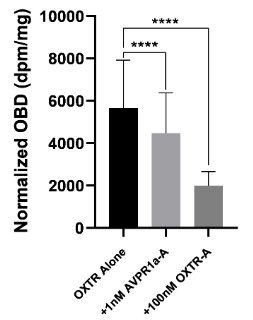
Differences in optical binding density (OBD) between conditions. Both competitors significantly reduced binding radioligand.
Figure 4
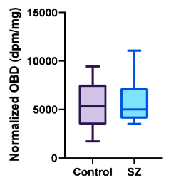
There is no significant difference in OXTR binding density between Control and SZ specimens.
Conclusions & Future Directions
- There was no detected variation in SN between SZ and control groups. Due to the complex anatomy, we were unable to look at the specific subregions of the SN, the pars reticulata (SNr), involved in inhibitory pathways, and pars compacta (SNc), involved in production of dopamine. Future studies might employ nissl staining to more clearly identify differences in OXTR between the subregions.
- The majority of specimen donors with SZ were taking antipsychotic medication. Not enough research has been performed considering antipsychotic medication on OXTR binding, so future studies may investigate these effects.
References
- Cloutier, M., Sanon Aigbogun, M., Guerin, A., Nitulescu, R., Ramanakumar, A. V., Kamat, S. A., DeLucia, M., Duffy, R., Legacy, S. N., Henderson, C., Francois, C., & Wu, E. (2016). The Economic Burden of Schizophrenia in the United States in 2013. The Journal of Clinical Psychiatry, 77(06), 764—761.
- Huey Yi Chong, Siew Li Teoh, David Bin-Chia Wu, Surachai Kotirum, Chiun-Fang Chiou, & Nathorn Chaiyakunapruk. (2016). Global economic burden of schizophrenia: a systematic review. Neuropsychiatric Disease & Treatment, 12, 357–373.
- Möller, H.-J. (2016). The Relevance of Negative Symptoms in Schizophrenia and How to Treat Them with Psychopharmaceuticals? Psychiatria Danubina, 28(4), 435–440.
- Meyer-Lindenberg, A., Miletich, R. S., Kohn, P. D., Esposito, G., Carson, R. E., Quarantelli, M., Weinberger, D. R., & Berman, K. F. (2002). Reduced prefrontal activity predicts exaggerated striatal dopaminergic function in schizophrenia. Nature Neuroscience, 5(3), 267–271.
- Montag, C., Brockmann, E.-M., Bayerl, M., Rujescu, D., Mueller (Müller), D. J., & Gallinat, J. (2012). Oxytocin and oxytocin receptor gene polymorphisms and risk for schizophrenia: A case-control study. The World Journal of Biological Psychiatry : The Official Journal of the World Federation of Societies of Biological Psychiatry, 14.
- Freeman SM, Walum H, Inoue K, Smith AL, Goodman MM, Bales KL, Young LJ. Neuroanatomical distribution of oxytocin and vasopressin 1a receptors in the socially monogamous coppery titi monkey (Callicebus cupreus). Neuroscience 2014; 273: 12–23.